Education
Webinars
The EUSOBI is proud to present a series of interactive, cased-based lectures, which aim at improving your clinical skills and answer your questions on breast imaging, from basic to advanced. The webinars are oriented to different levels of expertise, and provide worldwide interactive teaching on basic and advanced topics related to the diagnosis, staging and treatment of breast cancer. Each webinar is presented by one of our EUSOBI experts and we give you the opportunity to ask questions in our session.
Webinar places are limited and will be offered on a first-come, first-served basis. Early registration is recommended. The webinars are open for EUSOBI members only. Additionally, EUSOBI members will be able to view the webinar recording online at the EUSOBI website about 2-3 weeks after the webinar.
Webinar Series 1 // 2020 // Tips and Tricks in Breast MRI
The first series of webinars was entirely dedicated to the most complex and useful examination, Breast MRI. The experts were focusing on both technical and clinical issues and have addressed protocols, indications and reporting of breast MRI. Recordings of the webinars are available for EUSOBI Members.
Webinar Series 2 // 2021 // Advancements in Mammography
The second series of webinars is dedicated to Mammography. Experts will present the most novel x-ray-based imaging modalities clinically available for breast imaging and currently under research that could have a role in the future; offer balanced content between cutting edge science and good everyday practice; update on the role of different available modalities in screening; and discuss the role of artificial intelligence in screening and diagnosis of breast cancer.
- 2.1 / What is personalized screening? Francesca Caumo, Verona/IT
- 2.2 / The role of Digital Breast Tomosynthesis in screening and clinical practice Sophia Zackrisson, Malmö/SE
- 2.3 / Tomo-guided biopsy and Contrast-enhanced Tomosynthesis Daniela Bernardi, Milan/IT
- 2.4 / Contrast-enhanced Mammography: Technique and protocols Marc Lobbes, Sittard-Geleen/NL
>>> registration open - 2.5 / Contrast-enhanced Mammography: Clinical applications Fabienne Thibault, Paris/FR
- 2.6 / Contrast-enhanced Mammography: Experience from two centers – Session 1 Jonathan James, Nottingham/UK
- 2.7 / Contrast-enhanced Mammography: Experience from two centers – Session 2 Eva M. Fallenberg, Munich/DE
- 2.8 / Applications of AI in screening Karin Dembrower, Stockholm/SE & Valeria Romeo, Naples/IT
- 2.9 / Breast CT Magda Marcon, Zurich/CH
- 2.10 / Phase Contrast Mammography Marco Stampanoni, Villigen/CH
CME Accreditation
The EUSOBI Webinar Series 2 – Advancements in Mammography has been accredited by the European Accreditation Council for Continuing Medical Education (EACCME®) with 1 European CME credit (ECMEC®s) per webinar. Each medical specialist should claim only those hours of credit that he/she actually spent in the educational activity.
Through an agreement between the Union Européenne des Médecins Spécialistes and the American Medical Association, physicians may convert EACCME® credits to an equivalent number of AMA PRA Category 1 CreditsTM. Information on the process to convert EACCME® credit to AMA credit can be found at www.ama-assn.org/education/earn-credit-participation-international-activities.
According to UEMS regulations, attendees can only claim CME credits if the whole webinar has been attended. You will need to complete the event evaluation form after the webinar. As part of this process partcipants’ attendance will be monitored throughout the event. Only live-viewing can be accredited.
Next Webinar
MAR 24, 2021: EUSOBI Webinar 2.4 / Contrast-enhance Mammography: Technique and protocols
Speaker: Marc Lobbes, Sittard-Geleen/NL
Moderator: Thiemo van Nijnatten, Maastricht/NL
Date: Wednesday, March 24, 2021
Time: 17:00-18:00 CET
Registration is online here: https://attendee.gotowebinar.com/register/3285067378844985359
What you can expect from this webinar?
Contrast-enhanced mammography (CEM) is an upcoming breast imaging modality that relies on the injection of iodine based contrast agent and dual-energy mammography. The increased acceptance of CEM can be deducted from the increasing amount of installments and growing number of scientific publications. CEM indications might include: problem solving tool in equivocal findings during conventional mammography, preoperative staging, response monitoring in women treated with NAC, high risk screening, etc. However, it also starts with acquiring a robust CEM image. Hence, the current webinar will focus on the technical principles of CEM, patient handling and image acquisition. Some of the most common questions when initiating CEM within your breast center will be addressed, as well as the most common indications for CEM.
What can you learn during this webinar?
– Learn how to set up a CEM program in your hospital.
– Learn to acquire a high quality CEM image and know the technical principles behind it.
– Learn to cope with the most common artefacts and pitfalls.
– Discuss the most common indications for CEM.
Webinar Sponsors
Please CLICK HERE to find further details regarding our webinar sponsors.
Upcoming Webinars
APR 15, 2021: EUSOBI Webinar 2.5 / Contrast-enhanced Mammography: Clinical applications
Speaker: Fabienne Thibault, Paris/FR
Moderator: Daly Avendano, Monterrey/MX
Date: Thursday, April 15, 2021
Time: 17:00-18:00 CEST
MAY 12, 2021: EUSOBI Webinar 2.6 / Contrast-enhanced Mammography: Experience from two centers - Session 1
Speaker: Jonathan James, Nottingham/UK
Moderator: Mirjam Wielema, Groningen/NL
Date: Wednesday, May 12, 2021
Time: 17:00-18:00 CEST
MAY 26, 2021: EUSOBI Webinar 2.7 / Contrast-enhanced Mammography: Experience from two centers - Session 2
Speaker: Eva M. Fallenberg, Munich/DE
Moderator: Federica Leone, Milan/IT
Date: Wednesday, May 26, 2021
Time: 17:00-18:00 CEST
Past Webinars
FEB 25, 2021: EUSOBI Webinar 2.3 / Tomo-guided Biopsy and Contrast-enhanced Tomosynthesis
Speaker: Daniela Bernardi, Milan/IT
Moderator: Lucia Grana, Lugo/ES
Date: Thursday, February 25, 2021
Time: 17:30-18:30 CET
Recording
The recording will be available after 2-3 weeks.
FEB 9, 2021: EUSOBI Webinar 2.2 / The role of Digital Breast Tomosynthesis in screening and clinical practice
Speaker: Sophia Zackrisson, Malmö/SE
Moderator: Heike Preibsch, Tübingen/DE
Date: Tuesday, Feburary 9, 2021
Time: 17:00-18:00 CET
What can you expect from this webinar?
This lecture will cover the basics and advances of digital breast tomosynthesis, DBT, and its role in breast cancer screening and clinical breast imaging. Scientific background data will be presented, and cases will be used to illustrate pearls and pitfalls with DBT in different situations.
What can you learn during this webinar?
– the technique behind digital breast tomosyntesis, strengths and limitations
– the current scientific evidence and recommendations for use of DBT in screening
– clinical situations where DBT might be useful
Recording
The recording will be available after 2-3 weeks.
JAN 27, 2021: EUSOBI Webinar 2.1 / What is personalized screening?
Speaker: Francesca Caumo, Verona/IT
Moderator: Elisabetta Giannotti, Nottingham/UK
Date: Wednesday, January 27, 2021
Time: 17:00-18:00 CET
What can you expect from this webinar?
Decide to undergo screening is an individual choice. The main questions are: how large the benefit of screening is in terms of reduced breast cancer mortality and how substantial the harm is in terms of interval cancers (defined as a cancer diagnosed after a negative mammographic screen and before the next routine screen), reduction of sensibility of mammography in dense breast and overdiagnosis, which is defined as the detection of cancers with screening which would not have become clinically apparent in the woman’s lifetime in the absence of screening.
There are two strategies to improve the effect of a screening program: find a better first level test- Automated Breast Ultrasound System (ABUS)-Magnetic Resonance Imaging (MRI)-Digital Breast Tomosynthesis (DBT)-Hand-Held Ultrasound (HHUS) or try the way to overcoming effect of density and make a better risk classification to have different screening strategies including other imaging modalities, in addition to mammography, that might improve early detection of breast cancer.
International strategy (Wisdom Study; Dense Study, MyPeBs) is thinking about tailored screening modifying interval screen and using other modality.
The objective of the webinar is to describe this experience and show our project: RiBS (RiskBasedScreening)
Recording
Watch the webinar recording HERE
JUN 9, 2020: EUSOBI Webinar 1.8 / Clinical application: Management of MRI-only lesions
Speaker: Panagiotis Kapetas, Vienna/AT
Moderator: Mirjam Wielema, Groningen/NL
Date: Tuesday, June 9, 2020
Time: 18:00 CEST
What can you expect from this webinar?
MRI has the highest sensitivity for the identification of breast cancer among all imaging modalities. The increased implementation of breast MRI studies leads to the identification of a large number of additional lesions that necessitate further work-up. While some of these can be identified, characterized and/or biopsied through second-look ultrasound, a substantial number of these lesions are only evident in MRI and thus need to undergo an MRI-guided biopsy.
The aim of this webinar is to provide an insight into the management of breast lesions only visible on MRI. A thorough, focused second-look ultrasound examination is a prerequisite- after securing that the lesion has no sonographic correlate, an MRI-guided intervention is necessary. The procedure of MRI-guided biopsies and wire-localizations will be described. A thorough radiologic-pathologic correlation is extremely important, just like with any other image-guided intervention.
What can you learn during this webinar?
– Performance of second-look ultrasound
– Procedure of MRI-guided biopsies with different systems
– Pearls and pitfalls of MRI-guided biopsies
– Considerations on the congruence of the pathologic outcome
– Regulatory issues
Recording
Watch the webinar recording HERE
MAY 4, 2020: EUSOBI Webinar 1.7 / Clinical application: Screening
Speaker: Machteld Keupers, Leuven/BE
Moderator: Elisabetta Giannotti, Nottingham/UK
Date: Monday, May 4, 2020
Time: 17:00 CEST
What can you expect from this webinar?
Breast cancer is the most frequent cancer in women in Europe. Mammography is the only tool used in population based breast cancer screening in Europe that has proven to save lives while reducing aggressive treatment by detecting cancer at an earlier stage. A screening programme can only achieve those benefits if there is quality control, both for the screening modality as well as for all further diagnostic procedures and treatment.
This webinar aims to provide an overview of the literature and guidelines for breast cancer screening with MRI. Women at high risk for breast cancer due to genetic factors can be offered additional screening with ultrasound and MRI outside general population screening programmes. For women with average risk but with dense breasts, screening with MRI is not yet recommended by the European Guidelines. In order to meet the high quality standards of a good screening programme, several considerations have to be made.
What can you learn during this webinar?
– European guidelines on breast cancer screening and subsequent diagnosis.
– Criteria for quality control in breast cancer screening.
– The (dis)advantages of breast MRI in screening in a high risk population and in general population.
– The considerations regarding implementation of MRI in breast cancer screening for dense breasts.
– Case-based discussion of the pros and cons of breast MRI in screening.
Recording
Watch the webinar recording HERE
APR 14, 2020: EUSOBI Webinar 1.6 / Clinical application: Implant diagnostics
Speaker: Silvia Perez, Madrid/ES
Moderator: Daly Avendano, Monterrey/MX
Date: Tuesday, April 14, 2020
Time: 17:00 CEST
What can you expect from this webinar?
Nowadays, the treatment of breast cancer includes not only a safe treatment with the objective of having free margins, but also recover the shape of the breast. Besides, the augmented mammoplasty for esthetic reasons is more and more common. When the augmented mammoplasty is performed for reconstructive reasons is called heterologous reconstruction. The heterologous reconstruction uses external material to reconstruct the breast. The most frequent materials employed are the implants.
In this webinar we will discuss about the types of implants and also about the normal, abnormal appearance of them on MRI and possible complications.
What will you learn during this webinar?
– To know the different types of implants.
– To know the MRI protocol for the study of the implants.
– To know the normal appearance of the implants.
– To know the abnormal appearance of the implants.
– To know the signs of intracapsular rupture.
– To know the signs of extracapsular rupture.
– To know possible complications
– To know the possibility of recurrence
Recording
Watch the webinar recording HERE
MAR 24, 2020: EUSOBI Webinar 1.5 / Common issues and how to solve them
Speaker: Matthias Dietzel, Erlangen/DE
Moderator: Hajer Jarraya, Lille/FR
Date: Tuesday, March 24, 2020
Time: 17:30 CEST
Recording
Watch the webinar recording HERE
FEB 19, 2020: EUSOBI Webinar 1.4 / Problem solving or problem making?
Speakers: Pascal Baltzer, Vienna/AT; Paola Clauser, Vienna/AT
Date: Wednesday, February 19, 2020
Time: 18:00 CET
What can you expect from this webinar?
Breast MRI is commonly used in cases with equivocal or inconclusive findings on mammography, tomosynthesis and ultrasound. Breast MRI, though, might cause a non negligible number of additional biopsies and follow up for benign lesions. Thus, MRI is considered by many a problem maker, rather than a problem solver.
The literature and guidelines that discuss the topic will be presented.
The webinar will focus on:
– Defining problem solving breast MRI;
– Discussing the pros and contra of problem solving breast MRI;
– Giving example through cases on when MRI should (or should not) be used as a problem solving method.
Recording
Watch the webinar recording HERE
JAN 22, 2020: EUSOBI Webinar 1.3 / Optimize Diffusion Weighted Imaging
Speaker: Monique Dorrius, Groningen/NL
Wednesday, January 22, 2020
17:30 CET
What can I expect from this webinar?
Dynamic contrast enhanced magnetic resonance imaging is widely used in breast cancer imaging. From all breast imaging modalities MRI has the highest negative predictive value (> 98%), and therefore breast cancer can be exclude safely in case of a negative scan. However when during the assessment of DCE MRI enhanced breast lesions are found, most of the time differentiation between benign and malignant breast lesions is difficult. This is because of the considerable overlap in enhancement patterns between benign and malignant breast lesions. In general practice there is need for a second look ultrasound and probably an invasive procedure, like a core needle biopsy. In most of the cases the anxiety of the woman and the biopsy itself is unnecessary in benign lesions. To overcome the problem of equivocal enhanced breast lesions, the question arise whether Diffusion Weighted Imaging can play a role. If so, when and how should DWI be used in the diagnostic work up? What is an “optimal” DWI breast protocol? The answer on these questions will be addressed in this webinar. Furthermore, the effect of b value on the accuracy of breast DWI and the diagnostic performance of the region of interest methods are highlighted subjects. In the end there will be short comparison between the role of DWI and MR spectroscopy in the diagnostic workflow of breast lesions.
What will I learn during this webinar?
– The role of Dynamic Contrast Enhanced Magnetic Resonance Imaging in breast cancer imaging
– When to use Diffusion Weighted Imaging (DWI) in breast cancer imaging
– How to use Diffusion Weighted Imaging in breast cancer imaging; The “optimal” DWI protocol
– The effect of b-value on the diagnostic accuracy of breast DWI
– The diagnostic performance of different region of interest methods in DWI
– DWI versus MR spectroscopy
Recording
Watch the webinar recording HERE
DEC 12, 2019: EUSOBI Webinar 1.2 / Clinical application: Staging breast cancer
Speaker: Katja Pinker-Domenig, New York/US; Vienna/AT
Thursday, December 12, 2019
17:00 CET
What can I expect from this webinar?
Magnetic resonance imaging (MRI) of the breast is an established non-invasive imaging technique with several indications including preoperative staging of breast cancer. In breast cancer patients MRI of the breast may be performed to assess lesion extent, detect satellite lesions as well as other cancers in the ipsi- or contralateral breast. This presentation aims to provide a comprehensive overview on the possible indications of preoperative MRI, to discuss the potential advantages and disadvantages of preoperative MRI and to highlight the importance of thorough pre-examination information about MRI of the breast to be provided to cancer patients.
Recording
Watch the webinar recording HERE
NOV 4, 2019: EUSOBI Webinar 1.1 / Setting up your breast MRI protocol
Speaker: Pascal Baltzer, Vienna/AT
Monday, November 4, 2019
17:30 CET
What can I expect from this webinar?
A comprehensive review of what a standard clinical protocol should contain. Practical tips and typical pitfalls will be addressed. The participant should be able to set up a multiparametric breast MRI protocol after having attended this webinar.
What will I learn during this webinar?
- The basic sequences a clinical breast MRI protocol should contain;
- Tips and tricks to achieve robust image quality;
- Practicalities regarding an optimized workflow including patient care and positioning, contrast injection and sequence timing.
Recording
Watch the webinar recording HERE
Webinar Recording
You have missed our webinar(s)? We are happy to inform you that EUSOBI webinars are recorded and made available to our society members (after a period of approximately 1 month).
In order to access the recordings, please ensure your active membership and request your webinar password from office@eusobi.org.
Check available recordings HERE
Journal Club
Keep up-to-date with the current literature is essential for both residents and specialists, but it is not always easily doable. Many times, a critical appraisal of a study would be beneficial, but it is for many almost impossible.
That is why the EUSOBI Young Club decided to promote an online journal club with the aims of:
- disseminating new information in the field of breast diagnostic;
- promoting evidence-based medicine;
- teaching critical appraisal skills for reading and writing a scientific work;
- providing an opportunity for an interactive discussion with junior and senior experts.
Journal Club places are limited and will be offered on a first-come, first-served basis. Early registration is recommended. The Journal Club is open for EUSOBI members only. Additionally, EUSOBI members will be able to view the recording online at the EUSOBI website about 2-3 weeks after the Journal Club.
The discussants will focus on:
- presenting the structure and content on the paper;
- discussing the most relevant information for the clinical practice;
- discussing the other recent literature on the topic;
- sharing their experience.
Certificate of attendance / EUSOBI CME points
The EUSOBI Journal Club is accredited with 1 EUSOBI CME point per session! Only live-viewing can be accredited. Watching recordings will not be accredited.
In order to receive the certificate, the attendee has to complete the feedback survey which will be provided after the Journal Club. Furthermore, the attendance time must be longer than 45 minutes. These EUSOBI CME points count towards the EUSOBI diploma.
UPCOMING JOURNAL CLUB *BREAKING NEWS*
MAR 31, 2021: (Temporarily) change in Dutch breast cancer screening programme: Screening from 2 to 3 years
PRESENTER: Ruud Pijnappel, Utrecht/NL
DISCUSSANTS: Christiane K. Kuhl, Aachen/DE & Gabor Forrai, Budapest/HU
MODERATOR: Nisha Sharma, Leeds/UK
DATE: Wednesday, March 31, 2021
Time: 17:00-18:30 CEST
PAST JOURNAL CLUB
EUSOBI Journal Club No.3 / The Kaiser School discusses the DENSE trial
DISCUSSED ARTICLE: Supplemental MRI Screening for Women with Extremely Dense Breast Tissue
Bakker, M.F., de Lange, S.V., Pijnappel, R.M. et al., for the DENSE Trial Study Group. Supplemental MRI Screening for Women with Extremely Dense Breast Tissue. N Engl J Med 2019; 381:2091-2102. https://www.nejm.org/doi/full/10.1056/NEJMoa1903986
PRESENTER/MODERATOR: Mirjam Wielema, Groningen/NL
DISCUSSANTS: Pascal Baltzer, Vienna/AT; Matthias Dietzel, Erlangen/DE; Clemens Kaiser, Heidelberg/DE
DATE: Wednesday, December 9, 2020
TIME: 17:00 CET
RECORDING:
Watch the Journal Club recording HERE
Recordings are only available to EUSOBI Members in good standing!
EUSOBI Journal Club No.2 / International evaluation of an AI system for breast cancer screening
DISCUSSED ARTICLE: International evaluation of an AI system for breast cancer screening
McKinney, S.M., Sieniek, M., Godbole, V. et al. International evaluation of an AI system for breast cancer screening. Nature 577, 89–94 (2020). https://doi.org/10.1038/s41586-019-1799-6
PRESENTER: Nina Pötsch, Vienna/AT
DISCUSSANTS: Magda Marcon, Zurich/CH & Anton Becker, US
MODERATOR: Maria Adele Marino, Messina/IT
DATE: Monday, November 9, 2020
TIME: 17:30 CET
RECORDING:
Watch the Journal Club recording HERE
Recordings are only available to EUSOBI Members in good standing!
EUSOBI Journal Club No.1 / Recommendations during the COVID-19 pandemic
DISCUSSED ARTICLE: Recommendations for triage, prioritization and treatment of breast cancer patients during the COVID-19 pandemic. Curigliano G, et al, Breast. 2020 Apr 16;52:8-16.
PRESENTER: Anna D’Angelo, Rome/IT
DISCUSSANTS: Valentina Iotti, Regio Emilia/IT; Joao Horvat, Sao Paulo/BR
MODERATOR: Thiemo van Nijnatten, Maastricht/NL
DATE: Tuesday, June 23, 2020
TIME: 19:00 CEST
RECORDING:
Watch the Journal Club recording HERE
Recordings are only available to EUSOBI Members in good standing!
Journal Club Recording
You have missed the Journal Club? We are happy to inform you that the EUSOBI Journal Club is recorded and made available to our society members (after a period of approximately 1 month).
In order to access the recording, please ensure your active membership and request your personal password from office@eusobi.org.
EUSOBI Young Researcher Grant
One of the aims of EUSOBI is to support young researchers. To achieve this aim, the EUSOBI decided to support a maximum of 3 research grants every year, starting in 2021. These grants will be awarded to young researchers, who want to develop a project in the field of breast imaging.
ELIGIBILITY CRITERIA
- EUSOBI member in good standing below the age of 35;
- All projects should be mentored by a senior EUSOBI member who will be in charge of overall supervision of the project (preferably from the scientific committee, as an alternative a senior researcher in breast imaging);
- Each project should have a maximum length of one year (six additional months may be requested)
- The chief of the department where the work is to be performed should provide confirmation that the young researcher will be allowed to perform the research and that mentoring will be provided when needed.
The project will be evaluated by a selected Committee and based on:
- Originality (10 points) 22,3%
- Methodology and scientific validity (15 points) 33,3%
- Importance of anticipated results and transferability to clinical practice (15 points) 33,3%
- Previous experience on the topic (i.e. previous papers published) (5 points) 11,1%
Final works will be presented at the first EUSOBI Annual Scientific Meeting available.
Each paper that will be published using data from the research will include the following phrase among acknowledgments: “This research was partially/totally funded by the Young Researchers Grant awarded by the European Society of Breast Imaging”.
FUNDING
EUSOBI will devolve the sum of up to EUR 7.500 to this initiative divided into max three grants of EUR 2.500 each. The sum will be awarded directly to the EUSOBI member and not to his/her department/university.
Grants will be transferred to recipients with the following timescales:
- 40% when the research begins, as communicated by grant recipient;
- 30% after six months activity, upon presentation of interim results;
- 30% when the research will be published in a peer-review journal.
DEADLINE
EUSOBI will open a call at ECR 2021, with deadline by June 30th, 2021
- Applications will be rated by a jury, before September 2021
- The winner will be announced at the EUSOBI Annual Meeting 2021 (online)
APPLICATION
Projects should be written on the two forms provided.
Form A includes the title of the project, keywords, background, rationale, objectives, methods, transferability, impact of results and expected project timeline.
Form A must not include any reference to the names of the Young Researcher or Mentor, or any reference to previously published papers by the applicant or applicant’s group. Failure to comply with that will result in exclusion from consideration for the Grant.
Download Form A
Form B includes the title of the project, name and CV of the Young Researcher and name and CV of the Mentor. Additionally, we ask for 3 signatures.
Download Form B
Forms A and B should be saved in PDF file format and named as Form_A_[titleoftheproject] and Form_B_[titleoftheproject], respectively, and sent to the EUSOBI Office (office@eusobi.org).
Publications
COVID-19 related recommendations by EUSOBI
EUSOBI recommendations for breast imaging and cancer diagnosis during and after the COVID-19 pandemic
In this document the European Society of Breast Imaging (EUSOBI) provides recommendations for breast care provision and procedural prioritization at the times of the COVID-19 pandemic, being aware that medical decisions must be taken balancing individual and community safety as well as the safety of healthcare workers.
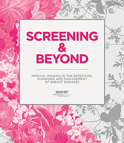
Published book
We are happy to announce the book, ‘Screening & Beyond’, created especially for the International Day of Radiology.
It is packed with information provided by many of the world’s top experts on breast imaging, including member of EUSOBI – European Society of Breast Imaging, the Society of Breast Imaging, ESTRO, European Institute for Biomedical Imaging Research and EUROPA DONNA – The European Breast Cancer Coalition
Published papers
Breast ultrasound: recommendations for information to women and referring physicians by the European Society of Breast Imaging
Andrew Evans, Rubina M. Trimboli, Alexandra Athanasiou, Corinne Balleyguier, Pascal A. Baltzer, Ulrich Bick, Julia Camps Herrero, Paola Clauser, Catherine Colin, Eleanor Cornford, Eva M. Fallenberg, Michael H. Fuchsjaeger, Fiona J. Gilbert, Thomas H. Helbich, Karen Kinkel, Sylvia H. Heywang-Köbrunner, Christiane K. Kuhl, Ritse M. Mann, Laura Martincich, Pietro Panizza, Federica Pediconi, Ruud M. Pijnappel, Katja Pinker, Sophia Zackrisson, Gabor Forrai, Francesco Sardanelli, for the European Society of Breast Imaging (EUSOBI), with language review by Europa Donna – The European Breast Cancer Coalition
Insights Imaging (2018). https://doi.org/10.1007/s13244-018-0636-z
A survey by the European Society of Breast Imaging on the utilisation of breast MRI in clinical practice.
Clauser P, Mann R, Athanasiou A, Prosch H, Pinker K, Dietzel M, Helbich TH, Fuchsjäger M, Camps-Herrero J, Sardanelli F, Forrai G, Baltzer PAT.
Eur Radiol. 2017 Nov 22. doi: 10.1007/s00330-017-5121-4
PMID:29168005
Mammography: an update of the EUSOBI recommendations on information for women.
Sardanelli F, Fallenberg EM, Clauser P, Trimboli RM, Camps-Herrero J, Helbich TH, Forrai G; European Society of Breast Imaging (EUSOBI), with language review by Europa Donna–The European Breast Cancer Coalition.
Insights Imaging. 2017 Feb;8(1):11-18. doi: 10.1007/s13244-016-0531-4. Epub 2016 Nov 16.
PMID:27854006
Translations: Estonian, Hungarian, Lithuanian, Polish, Portuguese, Serbian, Spanish, Turkish
Position paper on screening for breast cancer by the European Society of Breast Imaging (EUSOBI) and 30 national breast radiology bodies from Austria, Belgium, Bosnia and Herzegovina, Bulgaria, Croatia, Czech Republic, Denmark, Estonia, Finland, France, Germany, Greece, Hungary, Iceland, Ireland, Italy, Israel, Lithuania, Moldova, The Netherlands, Norway, Poland, Portugal, Romania, Serbia, Slovakia, Spain, Sweden, Switzerland and Turkey.
Sardanelli F, Aase HS, Álvarez M, Azavedo E, Baarslag HJ, Balleyguier C, Baltzer PA, Beslagic V, Bick U, Bogdanovic-Stojanovic D, Briediene R, Brkljacic B, Camps Herrero J, Colin C, Cornford E, Danes J, de Geer G, Esen G, Evans A, Fuchsjaeger MH, Gilbert FJ, Graf O, Hargaden G, Helbich TH, Heywang-Köbrunner SH, Ivanov V, Jónsson Á, Kuhl CK, Lisencu EC, Luczynska E, Mann RM, Marques JC, Martincich L, Mortier M, Müller-Schimpfle M, Ormandi K, Panizza P, Pediconi F, Pijnappel RM, Pinker K, Rissanen T, Rotaru N, Saguatti G, Sella T, Slobodníková J, Talk M, Taourel P, Trimboli RM, Vejborg I, Vourtsis A, Forrai G.
Eur Radiol. 2017 Jul;27(7):2737-2743. doi: 10.1007/s00330-016-4612-z. Epub 2016 Nov 2.
PMID:27807699
Translations: Estonian, Lithuanian, Polish, Portuguese, Serbian, Spanish, Turkish
Breast MRI: EUSOBI recommendations for women’s information.
Mann RM, Balleyguier C, Baltzer PA, Bick U, Colin C, Cornford E, Evans A, Fallenberg E, Forrai G, Fuchsjäger MH, Gilbert FJ, Helbich TH, Heywang-Köbrunner SH, Camps-Herrero J, Kuhl CK, Martincich L, Pediconi F, Panizza P, Pina LJ, Pijnappel RM, Pinker-Domenig K, Skaane P, Sardanelli F; European Society of Breast Imaging (EUSOBI), with language review by Europa Donna–The European Breast Cancer Coalition.
Eur Radiol. 2015 Dec;25(12):3669-78. doi: 10.1007/s00330-015-3807-z. Epub 2015 May 23. Review.
PMID:26002130
Translations: Estonian, Lithuanian, Portuguese, Serbian, Spanish, Turkish
Mammography: EUSOBI recommendations for women’s information.
Sardanelli F, Helbich TH; European Society of Breast Imaging (EUSOBI).
Insights Imaging. 2012 Feb;3(1):7-10. doi: 10.1007/s13244-011-0127-y. Epub 2011 Oct 28.
PMID:22695994
Breast MRI: guidelines from the European Society of Breast Imaging.
Mann RM, Kuhl CK, Kinkel K, Boetes C.
Eur Radiol. 2008 Jul;18(7):1307-18. doi: 10.1007/s00330-008-0863-7. Epub 2008 Apr 4. No abstract available.
PMID:18389253
Guidelines from the European Society of Breast Imaging for diagnostic interventional breast procedures.
Wallis M, Tardivon A, Helbich T, Schreer I; European Society of Breast Imaging.
Eur Radiol. 2007 Feb;17(2):581-8. Review. Erratum in: Eur Radiol. 2007 Feb;17(2):589. Tarvidon, Anne [corrected to Tardivon, Anne].
PMID:17013595
Press release
October 2018: Record number of participants travel to Athens to attend the EUSOBI Annual Scientific Meeting 2018
October 2018 (ESR, Vienna) – Athens played host to the European Society of Breast Imaging (EUSOBI) Annual Scientific Meeting, which took place on 11-13 October and saw its highest level of participation since the society was founded. With over 900 in attendance, the congress is on the fast lane to become the universal leading meeting in breast imaging.
Held in a different location each year, this year’s meeting saw over 100 additional participants compared to last year’s congress in Berlin.
The annual meeting was organised in collaboration with the Hellenic Society of Breast Imaging and involved the active participation of many Greek radiologists, but also pathologists, breast surgeons, medical oncologists and radiation oncologists. These participants, as well as many other worldwide experts helped bring an exciting programme to audiences, with highlights including dedicated sessions on tomosynthesis in screening/diagnostic settings, artificial intelligence and evidence-based imaging, and advanced/multiparametric ultrasound. This year’s EUSOBI Gold Medal, an award introduced in 2014, was given to Professor Christiane K. Kuhl from Aachen/Germany.
Part of the meeting’s success can be attributed to its expanded programme. This year, EUSOBI extended the congress to 2.5 days, offering a variety of pre-congress courses. These courses included updates on case-based BI-RADS classification, interventional procedures and B3 lesions. In addition and for the first time, the EUSOBI Young Club Committee organised a post-congress special symposium dedicated to young radiologists with an interest in breast imaging and diagnostics.
EUSOBI Past-President, Professor Gábor Forrai from Budapest/Hungary, is delighted with the annual meeting’s growth over the course of his three-year presidency. ‘Each year, I am so happy to see more and more people taking an interest in breast imaging. The fact that they are willing to give their time to come teach, learn and share ideas with colleagues here in Athens is an impressive mark of how dedicated those working in this field are’. During the congress in Athens Doctor Julia Camps Herrero from Valencia/Spain took over the presidency for the next two years. She will be taking office at an exciting time for the society, with EUSOBI having just reached a milestone of serving over 1,000 members for the first time since it was founded.
The European Society of Radiology (ESR) would like to congratulate the European Society of Breast Imaging and the Hellenic Society of Breast Imaging for the success of the meeting and look forward to seeing more great things from EUSOBI in the future.
You can learn more about EUSOBI’s annual meeting here.
September 22, 2016: EUSOBI Annual Scientific Meeting to kick off in Paris tomorrow
Thursday, September 22, 2016 (EUSOBI) – The European Society of Breast Imaging (EUSOBI) will hold its Annual Scientific Meeting in Paris, La Villette, in collaboration with the Society of Female Imaging – Société d’Imagerie de la Femme (SIFEM) on September 23–24, 2016. A breast magnetic resonance imaging (MRI) course for radiologists is also being held in advance on September 21–22.
Some 660 registered participants are expected to attend this year’s Annual Meeting, confirming a continuously increasing number of attendees over the years.
EUSOBI offers a balanced programme with cutting-edge science and good everyday practice. The Paris meeting will be a perfect opportunity to meet and exchange knowledge with experts and breast radiologists, as well as related companies from all over Europe and beyond.
The EUSOBI National Societies Network is made up of 29 countries. The first position paper from this large group of breast radiology societies will be published in European Radiology in October 2016, addressing the fundamental role of screening mammography for reducing the breast cancer mortality. EUSOBI unequivocally recommends all women to attend screening, according to their countries’ protocols.
This year, the 30th anniversary of contrast-enhanced breast MRI (1986–2016) will be celebrated with the EUSOBI Gold Medal Lecture delivered by Prof. Sylvia Heywang-Köbrunner, who was the first to report on this excellent diagnostic method in 1986. The Keynote Lecture will be delivered by Prof. Hedvig Hricak (Chairman of the Department of Radiology, Memorial Sloan-Kettering Cancer Center, New York, USA) on oncologic imaging in clinical decision making. The President of the U.S. Society of Breast Imaging, Prof. Elizabeth Morris (Chief of Breast Imaging at the Memorial Sloan-Kettering Cancer Center, New York, USA), will present the latest research and achievements in metastatic breast cancer.
The ‘Best way to screen’ session will tackle a hot topic as the debate on breast screening continues while the ‘Cutting edge’ and ‘B3 management’ sessions will include the latest scientific achievements. EUSOBI is currently focusing on breast MRI as this method already plays a major role in daily care, therapy planning and determining disease prognosis.
The EUSOBI Young Club will welcome its new active members at the special young scientists’ session, where the Young Investigator Award will be handed over and the best submitted poster abstracts will be presented.
EUSOBI will announce its new ‘5 points against breast cancer’ in Paris:
- In Europe, there are slightly different breast screening protocols in every country, but one statement is uniform: mammography saves lives and life quality – it can decrease breast cancer death by up to 40%.
- One in five breast cancers occur in women below 50, so screening should start before this age, at least at 45.
- 45% of breast cancers and related deaths occur in women at 65 and over. One in three breast cancers occur in women over 70, so screening should not stop at this age, but continue at least up to 75.
- If a woman has breast symptoms, the breast radiologist is the doctor to meet – he/she integrates all diagnostic techniques in order to give the most accurate information on breast health status e. g. cancer disease.
- The woman’s risk profile matters: personalised screening protocols may include ultrasound and/or MRI – but should always be interpreted together with mammography.
Recommended articles
Digital breast tomosynthesis (DBT): a review of the evidence for use as a screening tool
Gilbert FJ, Tucker L, Young KC
Clinical Radiology. 2016 Feb;71(2):141-50. doi: 10.1016/j.crad.2015.11.008
CONCERNS ABOUT SAFETY OF GADOLINIUM-BASED CONTRAST AGENTS (GBCAs)
A controversial debate about the safety of GBCAs recently prompted the European Medicines Agency and the U.S. Food and Drug Administration (see documents: FDA, EMA, ISMRM), to consider the clinical use of GBCAs in MRI. EUSOBI consider that indications for contrast-enhanced breast MRI as defined in our recommendations (Breast MRI: guidelines from the European Society of Breast Imaging and Breast MRI: EUSOBI Recommendations for women’s information) are still valid, and we believe that no breast MRI examination should be cancelled because the advantages (diagnosing breast cancer) clearly outweigh the possible problems contrast material could cause. We will modify our approach in the light of new evidence if appropriate.
Guidelines
ESR European Training Curriculum for Subspecialisation in Radiology
Ask the expert
Do you have a question on breast cancer?
Ask an expert via the EUSOBI Homepage! Our panel of experts in all the fields of breast will answer all of your questions. Share your thoughts with us on either breast imaging techniques, imaging interpretation, diagnosis, treatment, follow up or on research issues.
We are collecting your questions and publish them right here:
Can I find any information about EU standards for Breast Unit on the EUSOBI website?
Please find here the link to the EUSOBI Position Paper: https://pubmed.ncbi.nlm.nih.gov/27807699/
Additionally, this could be also helpful: https://www.sciencedirect.com/science/article/pii/S0960977620300606?via%3Dihub
answered by P. Clauser
At what age would you consider a women as a high risk candidate for developing breast cancer if you only take into account family history? By this, I am referring only to close relatives of the hypothetical candidate (ie. mother, sister, daughter). At what age is a woman thought of as high risk if she has one close family member with breast cancer? At what age is a woman thought of as high risk if she has 2 close family members with breast cancer?
There are specific tools to assess the risk of developing breast cancer, which do not only consider the family history of breast cancer or other risk factors. It is important to follow as much as possible well established guidelines and recommendations when defining a woman’s risk level.
Information on how to classify a patient as FH patient can be found here, according with NICE guidelines: https://cks.nice.org.uk/topics/breast-cancer-managing-fh/management/breast-cancer-managing-fh/
Starting age is influenced by different factors. Information from the High Risk screening programme in UK can be found here:
https://www.gov.uk/government/publications/breast-screening-higher-risk-women-surveillance-protocols
answered by E. Giannotti & P. Clauser
Is MRI indicated in patients >80 with bilateral breast cancer?
YES:
If surgical candidate with either dense breast/ suspicion of multifocality/-centricity on conventional imaging (with equivocal findings on conventional imaging).
If neoadjuvant chemotherapy planned
If not surgical candidate and neoendocrine treatment planned if MRI is tolerated (otherwise US)
NO:
Bilateral mastectomy planned
Non-dense breast with unifocal cancer/ no suspicion of multifocality/-centricity on conventional imaging
Obviously, in case of contraindications to MRI or contrast agent administration
answered by R. Mann, K. Pinker-Domenig and P. Clauser
A focus or focal area of NME on MRI breast that is separate to the known primary cancer in the context of a patient with known breast cancer, how does one manage it?
In case of a focus (enhancing lesion < 5 mm), it is important to make sure that there is no correlate in the pre-contrast and in the T2-weighted images that might help with further characterization towards a small suspicious mass or benign lesion. In the absence of a correlate on T1 and T2 weighted sequence, the symmetry should be evaluated: diffuse, symmetric foci are in general expression of diffuse benign changes in the breast.
In case of a single focus or focal non mass enhancement, there is only a little number of findings specific for a benign diagnosis. Small intralesional cysts, high ADC values and homogeneous and persistent enhancement curves suggest a benign finding. In the clinical context of cancer staging, it is very important whether the additional finding may be oncologically relevant (thus, discuss the impact on the therapy with the team). In many cases, such findings represent B3 lesions which e.g. in case of adjuvant radiation therapy and antihormonal therapy may be oncologically well treated without dedicated surgery. It is a difficult and case-by-case decision to biopsy such lesions or not (thus, all imaging and clinical information should be considered).
answered by P. Baltzer and P. Clauser
Does linear-ductal non mass stippled enhancement always have to enhance in the direction of the nipple to be considered worrisome...or is it possible that this type of worrisome non mass enhancement can configure in a direction away from the nipple?
Linear enhancement is most frequently intraductal in origin, and consequently follows the ductal structure of the breast.
The term stippled however does no longer exist, as stippled enhancement has been grouped with background parenchymal enhancement.
In the area of non-mass enhancement you might encounter a focal area of enhancement or regional enhancement that does not conform to a ductal tree structure. These may still be worrysome as they may be caused by diffusely growing infiltrative tumors (e.g. lobulars). In most of the cases you will find more features pointing in the direction of cancer (i.e. exceptionally rapid enhancement and wash-out, edema, low adc values, etc.)
answered by R.M. Mann
BEST OF Posters
Have a look at the ‘best of’ EUSOBI Scientific Posters presented at the Annual Scientific Meetings from 2016 to 2018.
News
Submit breast related news and publications here.
Journals
EUSOBI has an affiliation with the following medical journals:

Fellowship
The rapid increase in the demand for imaging women with symptoms and for population screening means that there is a need to sufficiently train further young radiologists to render confident diagnosis in breast imaging and intervention using all modalities. This has led the European Society of Radiology (ESR) and the European Society of Breast Imaging (EUSOBI) to take the initiative in establishing an exchange programme for fellowships or subspecialisation training in breast imaging.
Application period for 2020
February 24 – March 16, 2020
For further information please refer to the ESOR website.
Aim
Breast Imaging is at the heart of modern management of breast disease which aims to make a clear cut diagnosis as promptly as possible to discharge women with benign disease and make a non operative diagnosis of cancer. The programme offers an opportunity to complement subspecialisation training in breast imaging or an existing structured fellowship programme, through exchange. During three months of training the trainee will be provided with intense modular training in breast imaging and will be supervised by a specialised tutor in a pre-selected, highly esteemed, academic training centre in Europe.
The programme is aimed at board-certified radiologists within the first three years after certification who desire to become radiologists with a subspecialist interest in breast imaging.
Number of available places
During 2020, three such programmes will be offered and organised through ESOR and the successful applicant will receive a grant jointly provided from ESR/ESOR and EUSOBI.
Eligibility
- This programme applies to radiologists within the first three years after certification, who desire to become subspecialist radiologists in breast imaging.
- Core training in clinical radiology in line with national training scheme and in accordance with ESR syllabus. This should include core experience in breast imaging.
- Applicants must be proficient in English.
- ESR and EUSOBI membership fees for 2019 must be settled.
Applicants who were successful in the past cannot be considered for a second time.
Duration of fellowship and funding
The training will start in the second half of the year and last for three months. The fellow will receive a grant (€3.500,-) upon completion of the training, delivery of a final report and submission of original flight tickets and accommodation receipts. The grant is intended to contribute in part to travel and accommodation expenses during the training period. ESOR cannot guarantee that the grant will cover these costs in full. During the period of the training the fellow is responsible for covering his/her expenses and his/her own health insurance. The training itself is offered for free. Please note that in particular cases administrative charges may arise, which are to be covered by the grant.
Programme structure
Based on a weekly training programme, the trainee will familiarise his/herself with all aspects of breast imaging and intervention by case-by-case hands-on teaching on routine clinical cases provided by an experienced staff. The trainee will design a personalised programme with his/her tutor to cover areas that will be most useful to his/her anticipated practice, ie referral-based imaging, screening, interventional or MRI. At the end of the training programme the trainee would be expected to have developed a sound knowledge in subspecialty breast disease on which further training and experience may be built-up.
In non-native English speaking training centres teaching would be in English, while major radiological conferences and reporting may be in the local language. Some knowledge of the local language may be an advantage.
The trainee will be able to observe clinical activities, but will not have direct patient care responsibilities.
It is the trainee’s responsibility to communicate with the centre regarding the details of the training and whether more responsibilities than observer status can be obtained. ESOR solely acts as facilitator and coordinator between the training centre and the trainee.
Certification
After successful completion of the three months training the trainee receives a certificate from ESR/ESOR and EUSOBI. In order to receive this, the fellow must present a written report about his/her work and activities during the programme.
Scholarship
Application period for 2020
February 24 – March 16, 2020
For further information please refer to the ESOR website.
Aim
The ESOR Visiting Scholarship Programme (Europe) offers qualified trainees the opportunity to get to know another training environment and to kick off an interest for subspecialisation in radiology. During three months of training the scholars will be provided with a structured, modular introduction to different subspecialties and will be supervised by a specialised tutor in a pre-selected, highly esteemed academic training centre in Europe.
Number of available scholarships
During 2020, up to 36 scholarships are available on different topics and will be realised in partnership with Bracco and ESR. One additional scholarship for Spanish residents in partnership with the Spanish Foundation of Radiology and three scholarships for Greek residents supported by the Hellenic Radiological Society will be offered. Furthermore, two of the available scholarships will be provided for residents from Georgia within the ESR Support Initiative.
Eligibility
- These scholarships apply to residents in their 3rd, 4th or 5th year of training in radiology who desire to get the benefit of a mentored modular training of a specific subspecialised topic in radiology.
- Completion of the training curriculum from at least the first two years of residency is required, verified by the head of the department. Competence in producing a radiological report and communicating with clinicians and patients is appreciated, as well as knowledge of the principles of administration and management applied to a clinical department with multi-disciplinary staff and high-cost equipment.
- Previous exposure in any field of special interest, including basic knowledge of imaging methods, indications for imaging and clinical expectations, is advantageous.
- Applicants must be proficient in English.
- ESR membership fees for 2019 must be settled.
Applicants who were selected for this programme or the Visiting Scholarship Programme USA in the past cannot be considered again.
Duration of scholarship and funding
The training will start in the second half of the year and last for three months. The scholar will receive a grant (€3.500,-) upon completion of the training, delivery of a final report and submission of original flight tickets and accommodation receipts. The grant is intended to contribute in part to travel and accommodation expenses during the training period. ESOR cannot guarantee that the grant will cover these costs in full. During the period of the training the scholar is responsible for covering his/her expenses and his/her own health insurance. The training itself is offered for free. Please note that in particular cases administrative charges may arise, which are to be covered by the grant.
Programme structure
The programme offers a structured modular training in a highly esteemed training centre. The trainee will have the opportunity to follow and participate in the core knowledge training programme through tutorials and lectures, followed by hands-on teaching on routine clinical cases and/or modality techniques and protocols, led by an assigned tutor.
In non-native English speaking training centres teaching would be in English, while major radiological conferences and reporting may be in the local language. Some knowledge of the local language may be an advantage.
The trainee will be able to observe clinical activities, but will not have direct patient care responsibilities.
It is the trainee’s responsibility to communicate with the centre regarding the details of the training and whether more responsibilities than observer status can be obtained. ESOR solely acts as facilitator and coordinator between the training centre and the trainee.
Certification
After completion of the training programme, the resident receives a certificate from ESR/ESOR. In order to receive this, the scholar must present a written report about his/her work and activities during the programme.